
The Challenge
While uncommon, bladder and ureteral injuries occur during surgery. Early detection provides both the patient and clinician the greatest opportunity for appropriate intervention allowing for positive clinical outcomes.
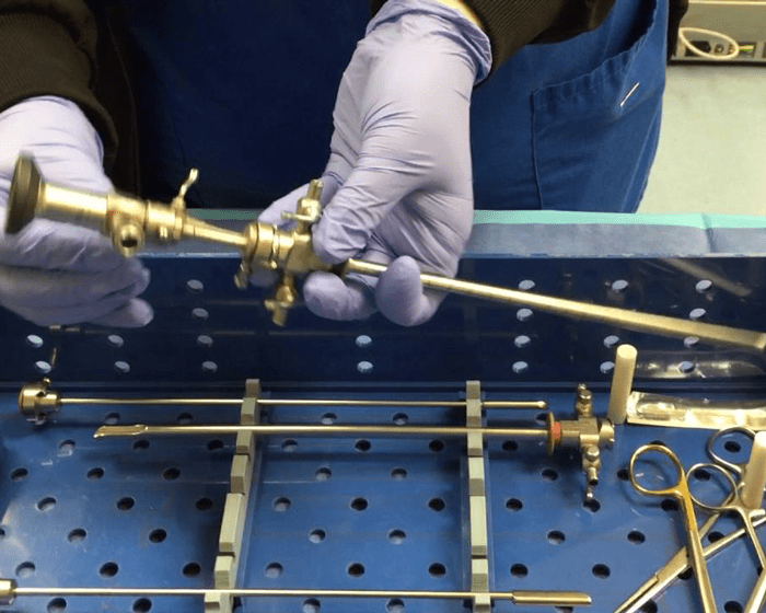
Cystoscopy, the insertion of a cystoscope through the urethra and into the bladder making it possible to visualize the inside of the bladder and ureteral flow, is one common practice employed to detect such injury. However, due to complex instrumentation, set-up, and availability of additional clinical support, this option is not always easily executed, and often only reserved for extreme circumstances.
Ureteral injury represents an uncommon but potentially morbid surgical complication...Iatrogenic ureteral injury increases the risk of hospital readmission and significant, potentially life threatening complications. Unrecognized ureteral injury markedly increases these risks, warranting a high level of suspicion for ureteral injury and a low threshold for diagnostic investigation.
While ureteral injuries are an uncommon surgical complication, it is estimated that 52-82% of iatrogenic injuries occur during gynecologic surgery…Colon and rectal procedures, such as low anterior resection (LAR) and abdominal perineal resection (APR) are responsible for 9% of all ureteral injuries.
Lower urinary tract injuries are a serious potential complication of laparoscopic hysterectomy. The risk of such injuries may be as high as 3%, and most, but not all, are detected at intraoperative cystoscopy…
Current Practices
To conduct a cystoscopy in surgical patients today requires multiple steps and involves several risks. Current cystoscopy techniques include:
- Insertion of a standard drainage catheter at the beginning of surgery
- Removal of the drainage catheter
- Assembly and insertion of the cystoscope sheath directly through the urethra
- Insertion of the cystoscope through the sheath
- Removal of the cystoscope
- Removal of the sheath
- Reinsertion of the drainage catheter
- Removal of the drainage catheter (when appropriate for patient post-op)
In summary a total of 8 passes into the bladder and 6 passes with contact against the urethra. Each of these steps takes time and may involve risks such as ureteral trauma, bladder perforation and an increased risk for urinary tract infections.

The CystoSure®
Advantage
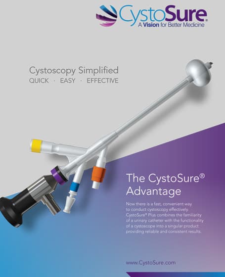
Now there is a fast, convenient way to conduct diagnostic cystoscopy effectively. Cystosure® Plus combines the familiarity of a urinary catheter with the functionality of a cystoscope into a singular product providing reliable and consistent results. The exclusive and patented pancake balloon and low profile tip positions scope for maximum viewing of the urethral ostia and bladder.
To conduct a cystoscopy in surgical patients with CystoSure® Plus simply:
- Insert the CystoSure® catheter
- Insert the CystoSure® cystoscope or compatible 2.9mm or smaller scope in hospital inventory
- Remove the scope
- Remove the catheter
In summary a total of 4 passes into the bladder and 2 passes against the urethra.


